
Live and Dried Blood Analysis with EMF Exposure
Challenge:
4-subject study Live blood analysis [1.2.3.4] involves visual examination of freshly-drawn capillary blood of subjects that is put between a glass cover slip and microscope slide and viewed using an optical microscope with video enhancement. It shows images of the various types of blood cells in their native state and plasma bodies as small as chylomicrons. It is regarded as an important assessment of the biological terrain of the body by holistic health practitioners.
The test requires either a dark-field or phase-contrast microscope and a high contrast videocamera attached to it. The magnification with video enhancement and projection on a monitor ranges up to 20,000X.

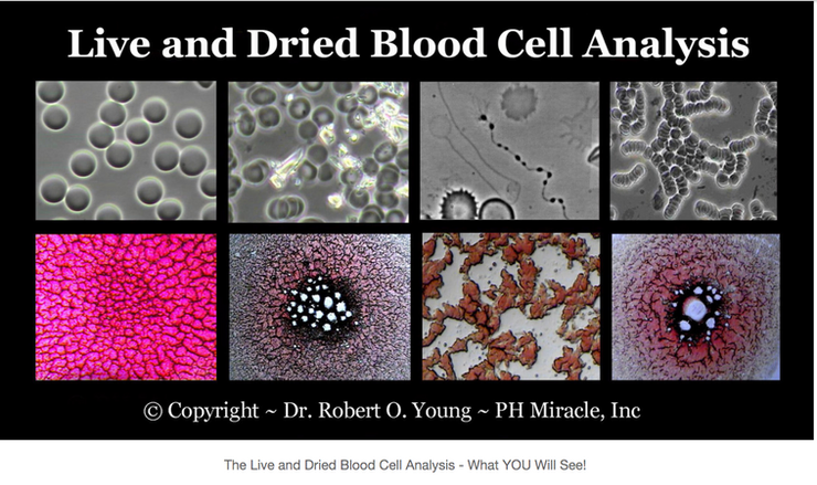
A remarkably detailed view of live, unstained blood is thus obtained, inaccessible by any other means. The size and shape of the cells, their morphological stability over time, states of agglutination, the presence of cell wall-deficient microbes[5] and the motility of white blood cells are just some of the parameters observed in video footage and microphotographs.
